


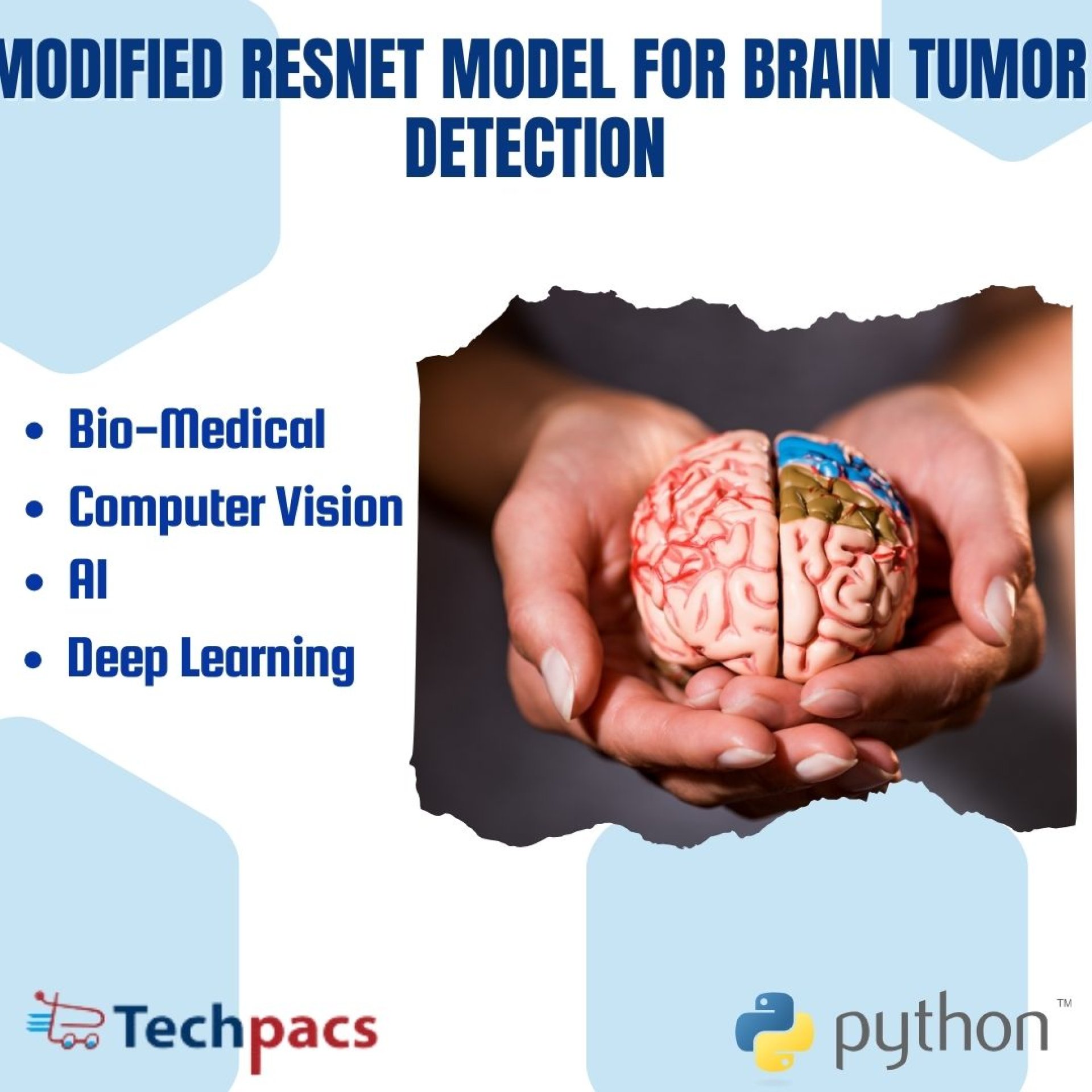
Innovative Approach in Brain Tumor Detection Using Combined T1 and T2 Modalities with ThinNet15 Framework
Problem Definition
After conducting a comprehensive literature review on AI-based systems for brain tumor detection and categorization, it is evident that several challenges hinder the effectiveness of existing systems. These challenges include network complexity, feature identification issues, and the refinement of medical images to improve accuracy. Additionally, most current systems are limited to utilizing a single type of image data, such as T1, T2, or FLAIR images, which contain different vital information. This limitation underscores the necessity for a more advanced system that can overcome these obstacles and provide a more efficient solution by incorporating information from multiple modalities, including T1, T2, FLAIR, and ADC images. Therefore, there is a pressing need to develop an innovative approach that can address the limitations of current AI-based systems and enhance the accuracy of brain tumor detection and classification by leveraging information from diverse image modalities while designing a less complex classification model.
Objective
The objective of the proposed work is to develop an innovative approach that can enhance brain tumor detection and categorization by utilizing information from images of multiple modalities, specifically T1 and T2. This approach aims to address the limitations of existing AI-based systems related to network complexity, feature identification issues, and image refinement, by incorporating information from diverse image modalities and designing a less complex classification model. The goal is to provide a more accurate and efficient solution for brain tumor diagnosis by preprocessing images, extracting features using a pretrained VGG network, combining features from multiple modalities, and utilizing a modified ResNet-34 model for brain tumor detection and classification. Additionally, leveraging techniques such as Gkmean segmentation, gaussian and bilateral filters for image enhancement, and the Kmean algorithm for segmentation, the proposed model seeks to overcome challenges in current AI-based systems and improve the accuracy and efficiency of brain tumor diagnosis.
Proposed Work
In this proposed work, the main objective is to develop an innovative approach that can enhance brain tumor detection and categorization by utilizing information from images of multiple modalities, specifically T1 and T2. The approach involves preprocessing the images to remove noise and segment the tumor section from both modalities. Feature extraction is then carried out using a VGG pretrained network, and the extracted features from both modalities are combined. Subsequently, a proposed network, based on a modified architecture of the ResNet-34 model, is utilized to simplify the model and improve its performance in detecting and classifying brain tumors. This approach aims to address the limitations of existing AI-based systems by offering a more accurate and efficient solution for brain tumor diagnosis.
The proposed approach leverages the Gkmean Segmentation technique for brain region segmentation, using gaussian and bilateral filters for image enhancement and the Kmean algorithm for segmentation. The VGG network is employed for feature extraction, while the ThinNet15 network is utilized for the classification task. By combining these techniques, the proposed model aims to overcome the challenges related to network complexity, feature identification, and image refinement faced by current AI-based systems. The rationale behind choosing these specific techniques and algorithms is to create a less complex classification model that can effectively handle images from multiple modalities and provide accurate results in brain tumor detection and categorization. This comprehensive approach offers a promising solution to enhance the accuracy and efficiency of AI-based systems in the field of medical image analysis for brain tumor diagnosis.
Application Area for Industry
This project can be utilized in various industrial sectors such as healthcare, medical imaging, pharmaceuticals, and biotechnology. The proposed solutions in this project can be applied within different industrial domains by addressing the challenges faced by existing AI-based systems in brain tumor detection and classification. Specifically, the system's ability to handle information from multiple modalities, including T1, T2, FLAIR, and ADC images, is crucial in the healthcare sector for accurate diagnosis and treatment planning. Moreover, the development of a less complex classification model can benefit medical imaging companies by streamlining the detection process and improving the efficiency of diagnosing brain tumors. The application of this project's proposed solutions in pharmaceuticals and biotechnology industries can lead to advancements in drug development and personalized medicine by providing more precise insights into brain tumor characteristics and behavior.
Overall, implementing these solutions can result in improved accuracy, efficiency, and effectiveness in detecting and categorizing brain tumors across various industrial sectors.
Application Area for Academics
The proposed project can enrich academic research, education, and training by introducing a novel approach to improve the detection and categorization of brain tumors using multiple modalities of imaging data. This unique methodology addresses the limitations of existing AI-based systems by incorporating information from both T1 and T2 modalities, along with a simplified classification model to enhance accuracy.
This research has the potential to contribute to advancing innovative research methods in the field of medical imaging analysis and AI. By utilizing algorithms such as Kmeans Clustering, Gaussian filter, Bilateral filter, and deep learning models like RESNET and VGG16, researchers, M.Tech students, and Ph.
D. scholars can explore new avenues for creating more effective solutions for brain tumor detection.
The relevance and potential applications of this project lie in its focus on bridging the gap between existing systems' complexity and limited scope in handling multiple types of imaging data. Researchers can benefit from the code and literature provided in this project to further their studies in medical image analysis, deep learning, and the application of AI in healthcare.
Future scope for this project includes expanding the research to include more modalities of imaging data, such as FLAIR and ADC images, to enhance the overall accuracy and efficiency of brain tumor detection systems.
Additionally, exploring the integration of other advanced algorithms and deep learning models can further improve the overall performance of the classification model.
Algorithms Used
Kmeans Clustering is used for image segmentation to separate the tumor section from both T1 and T2 images. Gaussian and Bilateral filters are applied for noise removal and smoothing of the images, enhancing the quality of the input data for subsequent processing.
Deep Learning models RESNET and VGG16 are utilized for feature extraction from the preprocessed images. The extracted features from both modalities are combined to capture a comprehensive representation of the tumor characteristics, leading to better detection and classification results.
The proposed ResNet-34 based network is modified to simplify the model and improve its performance specifically for brain tumor detection and classification tasks.
This modified architecture aims to address the limitations of existing AI-based systems, providing a more accurate and efficient solution for identifying and categorizing brain tumors.
Keywords
SEO-optimized keywords: Brain tumors, medical imaging, MRI, CT, PET, early detection, automated diagnosis, machine learning, transfer learning, data augmentation, ensemble learning, image variability, small dataset size, inter-observer variability, computational complexity, noise removal, segmentation, feature extraction, VGG pretrained network, ResNet-34 model, brain tumor detection, brain tumor classification, multiple modalities, T1 images, T2 images, FLAIR images, ADC images, classification model, network complexity, accurate detection, innovative approach, brain images, classification tasks.
SEO Tags
Brain tumors detection, brain tumor classification, AI-based systems, medical imaging, MRI, CT images, PET imaging, early detection of brain tumors, automated diagnosis, machine learning in medical imaging, transfer learning for brain tumor detection, data augmentation in medical image analysis, ensemble learning for brain tumor classification, challenges in brain tumor detection, refining medical images, brain tumor segmentation, feature extraction in brain tumor detection, ResNet-34 model, VGG pretrained network, brain tumor classification model, improving accuracy in brain tumor detection, research on brain tumor detection, brain tumor treatment, non-invasive imaging techniques, computational complexity in medical imaging, noise removal in MRI images, small dataset size in medical imaging, inter-observer variability in brain tumor detection, ThinNet15 classification network, image classification for brain tumors, multi-modality imaging in brain tumor detection, PHD research topic, MTech research project, Brain tumor research framework.
| Shipping Cost |
|
No reviews found!


No comments found for this product. Be the first to comment!