


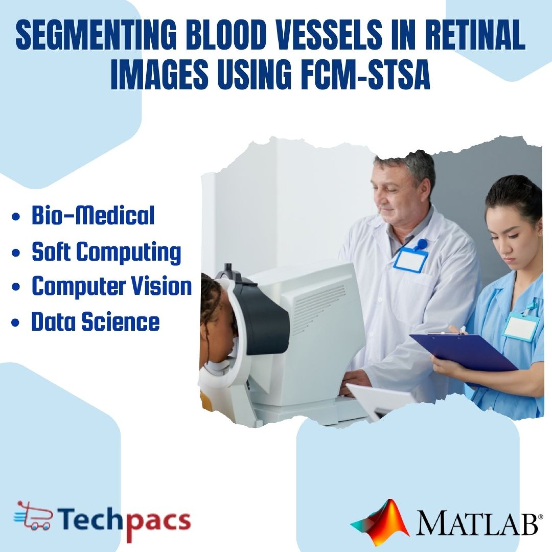
Enhancing Retinal Blood Vessel Segmentation Using FCM-STSA Algorithm and Image Enhancement.
Problem Definition
The literature review of automated retinal blood vessel extraction methods reveals several key limitations and challenges that need to be addressed. One of the primary issues identified is the difficulty in obtaining the Region of Interest (ROI) from retinal images, as the complex structure poses a challenge for researchers. This limitation hinders the accuracy of current segmentation models and results in high computational time, as the data processing is slow. Additionally, factors such as noise and lighting conditions further degrade the efficacy of the analysis techniques, leading to poor quality images and low accuracy rates. Another crucial area that has been overlooked is the lack of focus on enhancing image quality during the pre-processing phase, which can also contribute to subpar results.
These limitations underscore the necessity for developing an effective approach that not only overcomes the existing challenges but also enhances the accuracy of blood vessel detection rates in retinal images.
Objective
The objective of the proposed model is to address the limitations and challenges faced by existing retinal blood vessel segmentation methods. This is achieved by focusing on image enhancement and segmentation to improve the accuracy of detection rates. The model aims to extract retinal blood vessels more effectively and efficiently from images by enhancing their quality before applying segmentation techniques. By utilizing techniques such as Adaptive Histogram Equalization (AHE) for image enhancement and Sine Tree-Seed Algorithm (STSA) and FCM clustering for segmentation, the model seeks to overcome issues such as noise, irregular lighting, and degraded image quality that affect current segmentation models. Through a systematic approach involving data collection, image enhancement, and segmentation techniques, the objective is to demonstrate significant improvements in the accuracy of detecting retinal blood vessels, thereby enhancing the overall segmentation process and improving the detection rate of blood vessels in retinal images.
Proposed Work
In this work, the proposed model aims to address the limitations and challenges faced by existing retinal blood vessel segmentation methods. By focusing on image enhancement and segmentation, the model strives to improve the accuracy of detection rates. The main objective is to extract retinal blood vessels more effectively and efficiently from images by enhancing their quality before applying segmentation techniques. To achieve this, the model goes through various phases such as data collection, layer extraction, ROI extraction, image enhancement, segmentation, and performance evaluation. The use of Adaptive Histogram Equalization (AHE) technique is instrumental in improving the quality of images by mitigating the effects of noise, irregular lighting, contrast, and other factors.
The enhanced images are then subjected to segmentation using Sine Tree-Seed Algorithm (STSA) and FCM clustering approach to achieve optimal results. The segmented images from different techniques are combined to demonstrate the efficacy of the proposed approach.
By implementing a hybrid of FCM clustering algorithm and STSA optimization algorithm for the segmentation of blood vessels in retinal fundus images, the proposed model aims to enhance the accuracy and efficiency of the retinal blood vessel segmentation process. The model addresses the research gap identified in the literature survey by focusing on overcoming the limitations of current models, such as high computational time, difficulty extracting the Region of Interest (ROI), and degraded image quality leading to lower accuracy rates. The rationale behind choosing the specific techniques lies in their effectiveness in improving the quality of images and accurately segmenting retinal blood vessels.
Through a systematic approach involving data collection, image enhancement, and segmentation techniques, the proposed model aims to demonstrate significant improvements in the accuracy of detecting retinal blood vessels. The choice of algorithms and technology, therefore, serves the purpose of achieving the objective of enhancing the segmentation process and ultimately improving the detection rate of retinal blood vessels.
Application Area for Industry
This project can be used in various industrial sectors such as healthcare, agriculture, manufacturing, and security. In the healthcare sector, the accurate extraction of retinal blood vessels can aid in the early detection and monitoring of diseases like diabetes and hypertension. In agriculture, this project can help in analyzing plant health and growth by studying the blood vessels in plant leaves. In manufacturing, the precise segmentation of blood vessels can be utilized for quality control purposes and in security, it can be used for biometric authentication systems. The proposed solutions of enhancing image quality and utilizing advanced segmentation techniques can provide industries with more accurate and efficient results.
By improving the accuracy of retinal blood vessel extraction, industries can benefit from enhanced diagnostic capabilities, better decision-making processes, and improved overall performance.
Application Area for Academics
The proposed project on retinal blood vessel segmentation aims to enrich academic research, education, and training in the field of medical image analysis. By addressing the limitations of existing models and focusing on image enhancement and segmentation techniques, this project offers a valuable contribution to the advancement of innovative research methods and data analysis within educational settings.
The relevance of this project lies in its potential applications for researchers, MTech students, and PhD scholars in the field of medical imaging and computer vision. The code and literature developed for this project can be utilized by researchers to enhance their understanding of retinal blood vessel segmentation, improve the accuracy of detection rates, and explore new techniques for image enhancement and segmentation. MTech students can leverage the project's methodologies and algorithms to gain practical experience in implementing advanced image processing techniques, while PhD scholars can utilize the findings for further research and experimentation in the domain.
The technologies covered in this project, such as Fuzzy C-Means clustering, Sine Tree-Seed Algorithm, Adaptive Histogram Equalization, and Average filters, offer a comprehensive toolkit for researchers and students to explore various approaches to retinal image analysis. By employing these advanced techniques, individuals can enhance their skills in data processing, segmentation, and evaluation of medical images, ultimately contributing to the development of impactful research in the field.
In the future, the scope of this project could be expanded to include real-time processing of retinal images, automation of segmentation techniques, and integration with machine learning algorithms for enhanced accuracy and efficiency. By continuing to innovate and refine the proposed model, researchers and students can make significant strides in the field of medical image analysis, paving the way for improved diagnostic tools and treatments for various eye-related diseases.
Algorithms Used
The proposed work focuses on enhancing the accuracy of retinal blood vessel segmentation using various algorithms. The image enhancement phase utilizes the Adaptive Histogram Equalization (AHE) technique to improve image quality by neutralizing the effects of noise, irregular lighting, and contrast. Subsequently, the images are divided into two categories for further processing. In one approach, the image is segmented after being divided into sub-parts, while in the other approach the enhanced image is directly segmented. The segmentation phase employs the Sine Tree-Seed Algorithm (STSA) and Fuzzy C-Means (FCM) clustering technique to extract retinal blood vessels effectively.
The segmented images from both approaches are then combined to assess the effectiveness of the proposed model in enhancing accuracy and efficiency in retinal blood vessel segmentation.
Keywords
SEO-optimized keywords: blood vessel segmentation, FCM-STSA method, retinal fundus images, image processing, image segmentation, medical image analysis, ophthalmology, retina, computer-aided diagnosis, feature extraction, fuzzy c-means (FCM), thresholding, image enhancement, image filtering, vessel detection, biomedical imaging, diabetic retinopathy, optical coherence tomography, medical imaging, artificial intelligence, ROI extraction, adaptive histogram equalization, DRIVE dataset, STARE dataset, segmentation techniques, Sine Tree-Seed Algorithm (STSA), image quality enhancement, computational time, noise reduction, lightening correction.
SEO Tags
Blood vessel segmentation, FCM-STSA method, Retinal fundus images, Image processing, Image segmentation, Medical image analysis, Ophthalmology, Retina, Computer-aided diagnosis, Feature extraction, Fuzzy C-Means, Thresholding, Image enhancement, Image filtering, Vessel detection, Biomedical imaging, Diabetic retinopathy, Optical coherence tomography, Medical imaging, Artificial intelligence
| Shipping Cost |
|
No reviews found!


No comments found for this product. Be the first to comment!